Brown patches on the face are often grouped under one word: pigmentation. Yet three common conditions sit behind most of them, and they behave differently. Melasma, sun spots (solar lentigines) and post-inflammatory hyperpigmentation (PIH) have different triggers, patterns and outlooks.
Knowing which one you may be dealing with helps you ask better questions at a dermatology appointment. It also explains why a treatment that clears one type of mark can make another worse.
This article is a guide to the differences, not a way to diagnose yourself. Any new, changing or unusual spot deserves a professional look.
Why they can look alike
All three involve extra melanin, the pigment that gives skin its color. In each case, pigment-producing cells (melanocytes) make more melanin, or melanin collects where it normally would not.
The differences lie in what drives that extra pigment and where in the skin it sits.
Melasma
What it looks like
DermNet describes melasma as blotchy, brownish patches on both sides of the face, usually with irregular borders. The patches are flat and cause no itching or pain. The British Association of Dermatologists (BAD) notes that the color can be brown or grayish.
Common patterns include:
- Centrofacial: forehead, cheeks, nose and upper lip. DermNet reports this pattern in 50 to 80% of cases.
- Malar: mainly the cheeks.
- Mandibular: along the jawline.
- Extrafacial: sun-exposed forearms, upper arms and shoulders.
Who tends to get it
Melasma is more common in women, and the American Academy of Dermatology (AAD) notes higher risk between roughly ages 20 and 40. It occurs more often in medium and darker skin tones. Family history is common: DermNet reports that about 60% of patients have affected relatives, and the AAD cites one study in which 48% of 324 patients reported an affected blood relative.
What drives it
The exact cause is not known. Recognized contributors include:
- Ultraviolet (UV) light from the sun, sunbeds and phototherapy
- Visible light, especially high-energy blue light, which BAD says may contribute (it adds there is no evidence that light from personal devices has an effect)
- Pregnancy, birth control pills and hormone replacement
- Some medications, including certain anti-seizure drugs
- Thyroid disease
Melasma tends to look stronger in summer and lighter in winter, according to BAD. It is not contagious and does not turn into skin cancer.
Sun spots (solar lentigines)
What they look like
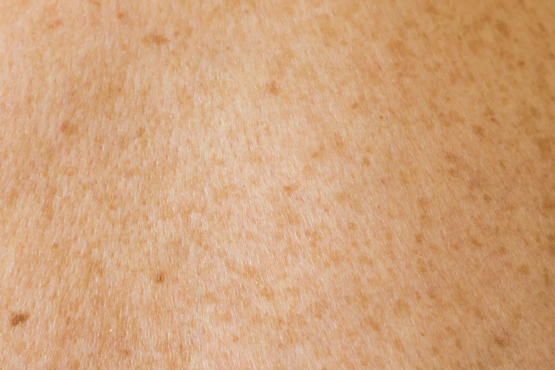
Solar lentigines are also called age spots, liver spots or sun spots. DermNet describes them as well-defined, usually round or oval spots that are flat or slightly raised. They range from yellow or tan to dark brown, and each spot is usually an even color. Size varies from a few millimeters to a few centimeters, and neighboring spots can merge.
They appear on areas with long-term sun exposure: the backs of the hands, forearms, upper body, shins and face.
Who tends to get them
They mostly affect middle-aged and older adults. DermNet notes they are most frequent in fair skin and also common in Asian populations, where they often appear on the face. Unlike freckles, they stay visible all year, though they may darken slightly in summer.
What drives them
The cause is chronic UV exposure, from sunlight or from sources such as sunbeds. Sunburn history and sunbed use are linked to higher risk.
Post-inflammatory hyperpigmentation
What it looks like
PIH is darkening that follows an injury or an inflammatory skin problem, such as a burn, dermatitis or an infection. DermNet explains that the patches appear in the same place as the original problem once it heals. They range from light brown to black. When pigment sits deeper in the skin, it can take on a gray, purple or brown tone.
Who tends to get it
Anyone can develop PIH, but it is more common in darker skin. In darker skin it also tends to be more intense and last longer.
What drives it
Inflammation stimulates melanocytes, and damage to the skin layers lets melanin collect in skin cells or the deeper dermis. Sun exposure can darken existing marks.
Side-by-side comparison
| Feature | Melasma | Sun spots | PIH |
|---|---|---|---|
| Typical shape | Blotchy patches, irregular borders | Well-defined round or oval spots | Follows the shape of the earlier injury or rash |
| Usual location | Both sides of the face, often symmetrical | Hands, forearms, face, upper body | Wherever inflammation occurred |
| Main triggers | UV, visible light, hormones, family history | Long-term UV exposure | Dermatitis, infection, burns, other skin injury |
| Seasonal change | Often worse in summer | Present year-round, may darken slightly | Can darken with sun |
| Typical age | Often starts between 20 and 40 | Middle age and older | Any age |
How a dermatologist tells them apart
Diagnosis is usually made by looking at the skin. DermNet lists two common tools:
- Dermoscopy: a magnifying device that shows pigment patterns. Sun spots show features such as a faint pigment network and well-defined or “moth-eaten” borders.
- Wood lamp: an ultraviolet lamp that can make epidermal (surface) melasma look more obvious.
A skin biopsy is occasionally done to rule out other conditions. This matters because some lesions can resemble benign spots. DermNet notes that atypical solar lentigines can be hard to tell apart from melanoma in situ, an early skin cancer. Spots with several colors, or ones that change, should be checked.
Depth matters
Both melasma and PIH can sit in the epidermis (upper layer), the dermis (deeper layer) or both. DermNet notes that epidermal melasma usually responds better to treatment, while dermal melasma usually responds poorly. Mixed melasma, the most common type, often improves partially.
Why the difference changes treatment
Treatment approaches overlap, but the priorities differ.
- Melasma usually needs a combination approach and long-term sun protection. DermNet cautions that procedures such as lasers and peels can worsen melasma or trigger new PIH. BAD notes there is no cure at present, and melasma often returns after treatment stops.
- Sun spots are harmless and need no medical treatment. When people choose to treat them for appearance, DermNet lists options such as cryotherapy, pigment-specific lasers, intense pulsed light, peels and topical agents.
- PIH often fades over time, though it can persist for a long time in darker skin. DermNet lists daily broad-spectrum sunscreen, camouflage and topical agents, usually in combination, and notes that physical treatments can make it worse by injuring the skin.
It is also possible to have more than one type at once. A person with melasma can develop PIH after a harsh peel, and sun spots can appear alongside either.
Questions worth bringing to an appointment
- Which type of pigmentation do I have, and is it in the upper or deeper skin?
- Do any of my spots need a closer look or a biopsy?
- Could a medication or hormone be contributing?
- Which treatments carry a risk of making my skin darker?
A dermatologist can examine your skin directly and suggest options that suit your skin type and history.
Sources
- Melasma (facial pigmentation) DermNet
- Solar lentigo DermNet
- Postinflammatory hyperpigmentation DermNet
- Melasma: Causes American Academy of Dermatology
- Melasma patient information leaflet (June 2024) British Association of Dermatologists
This article is general information, not medical advice. It does not replace an examination, and treatment for pigmentation should be planned with a dermatologist or another qualified clinician who has seen your skin.